Case: Cervical laminectomy for meningioma at the C2 level
This is a 71 year old woman presents with 10 months of progressive weakness of arms and legs. She started with difficulty walking and has progressed to the point that she needs significant assistance to walk. She also reports difficulty with voiding.
Her examination reveals severe left-sided weakness most pronounced in left hand and hip muscles. She is hyperreflexic.
- All
- Pre-Op
- Intra-op
- Post-op
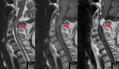 Sagittal MRI images (T2, T1, and T1 with contrast; left-to-right) showing a tumor (red arrow) behind the spinal cord at the level of C2. It enhances brightly with the administration of contrast.
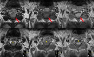 Axial MRI images (T2, T1, and T1 with contrast; left-to-right) showing a tumor (red arrow) behind the spinal cord at the level of C2. It is more prominent on the left side, it is compressing the spinal cord (yellow star (*)), and it enhances brightly with the administration of contrast.
 Sagittal (left) and axial (right) CT scan images showing the bony anatomy of the patient. Degenerative changes are notes, but the tumor is not visualized on these images.
 Axial CT scan images showing soft tissue including the tumor (red arrow).
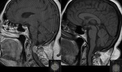 Midsagittal images from two brain MRI’s. The left images is from a recent MRI, and the right is from 12 years prior. Note the large tumor on the new MRI and the hint of a small lesion on the MR image on the right.
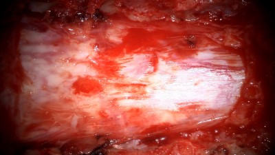 Intraoperative photo showing the covering of the spinal cord, the dura mater. The bone (lamina) over the dura has been removed. The dura has a normal appearance on its outer surface. Orientation: this is the back of the patient with the head to the left of the image and the legs to the right.
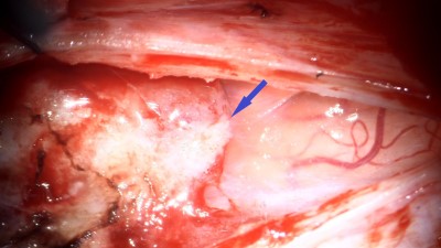 Intraoperative photo showing the tumor and the spinal cord. The covering of the spinal cord has been opened. The blue arrow points to the tumor which is on the left of the image. The spinal cord is to the right of the arrow and is covered by a translucent membrane, the arachnoid mater.
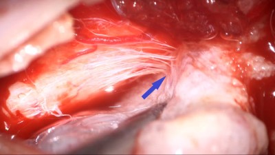 Intraoperative photo showing microdissection of the tumor from the spinal cord. The tumor is being retracted to the right, exposing many small bands of tissue (arrow) connecting the tumor to the spinal cord on the left.
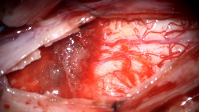 Intraoperative photo showing the dorsal surface (back side) of the spinal cord after the tumor has been resected.
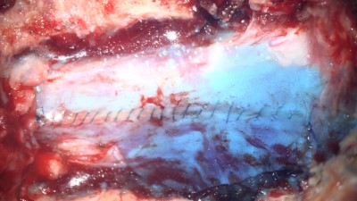 Intraoperative photo showing the dural repair. The dura has been sutured together, and the water-tight repair is reinforced with a thin layer of sealant which appears as a blue film.
 Postoperative sagittal MRI images (T2, T1, and T1 with contrast; left-to-right) showing postop changes without residual tumor 3 months after her surgery. Compare with preop MRI, Figure 1.
 Axial MRI images (T2, T1, and T1 with contrast; left-to-right) showing postop changes without residual tumor 3 months after her surgery. Compare with preop MRI, Figure 2.


