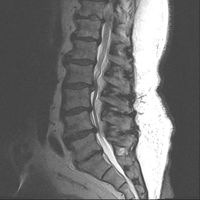
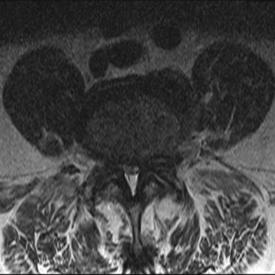
This is a 69 year old male who presents with severe right ankle weakness (foot drop). He has bilateral foot numbness and moderate back pain radiating into his legs. Neurodiagnostic testing confirms L5 radiculopathy.
  • All
  • Pre-Op
  • Intra-op
  • Post-op
Midsagittal T2 MRI image shows multiple levels of degeneration including L4/5. Alignment and lordosis are well maintained.
Axial T2 MRI image through the L4/5 level. It shows severe central and lateral recess stenosis from disc displacement as well as ligamentous and facet hypertrophy.
Intraoperative photo taken through the operating microscope. Cranial is to the left and caudal to the right. It shows that bone has been removed on both sides of the midline (aka bilateral hemilaminotomies; blue star) and the spinous processes are intact between.
Intraoperative image now shows that the a portion of the spinous processes and lamina have been removed in the midline (blue star) in preparation for the Coflex device.
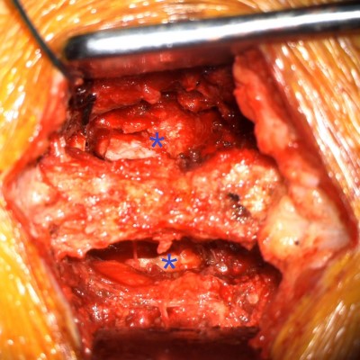
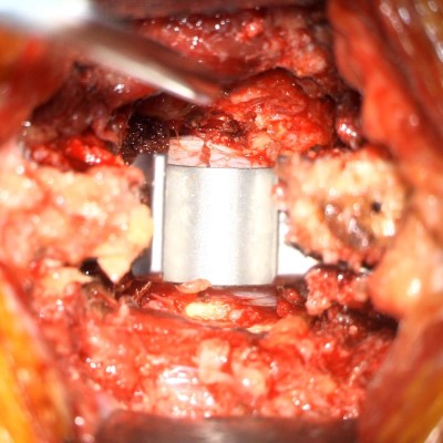
Intraoperative video showing the placement of a sizer preparing for placement of the Coflex device. Note how L4 (on the left) moves away from L5 (on the right) as the red sizer is placed in the laminar defect.
Intraoperative image showing the Coflex stabilization device in the laminar defect. Note the wide decompression on both sides of the device (above & below on image).
Postoperative x-rays of the lumbar spine in the AP (left) and lateral (right) planes. It shows the Coflex device in the L4/5 interspace.
4 months postop patient reports improvement of back and leg pain and increased right ankle strength.
Short video demonstrating how the Coflex device works
Click here for more info